


• Anti Hepatitis B Envelope Antibody (AHBe) - Total New
    935.00₹
    In stock
    All over India
    0 Reviews
    The **Anti-Hepatitis B Envelope Antibody (AHBe) - Total** test (also known as the Anti-HBe or HBeAb test) is a crucial blood marker used to monitor and evaluate a Hepatitis B virus (HBV) infection.

    This test detects total antibodies produced by the body’s immune system to target the Hepatitis B "e" antigen (HBeAg), a protein associated with active viral replication. A positive or reactive result generally signals "seroconversion"—meaning the immune system is gaining control, viral multiplication is slowing down, and the patient's infectivity (risk of transmitting the virus to others) has significantly decreased. Doctors use it alongside other HBV tests to determine the infection stage and assess treatment success.
    The **Anti-Hepatitis B Envelope Antibody (AHBe) - Total** test (also known as the Anti-HBe or HBeAb test) is a crucial blood marker used to monitor and evaluate a Hepatitis B virus (HBV) infection. This test detects total antibodies produced by the body’s immune system to target the Hepatitis B "e" antigen (HBeAg), a protein associated with active viral replication. A positive or reactive result generally signals "seroconversion"—meaning the immune system is gaining control, viral multiplication is slowing down, and the patient's infectivity (risk of transmitting the virus to others) has significantly decreased. Doctors use it alongside other HBV tests to determine the infection stage and assess treatment success.
    0 Comments 0 Shares 3K Views 0 Reviews
  • AFB-M.Tb detection (M.Tb/NTM Detection) by CBNAAT BAL New
    2,700.00₹
    In stock
    Hyderabad
    0 Reviews
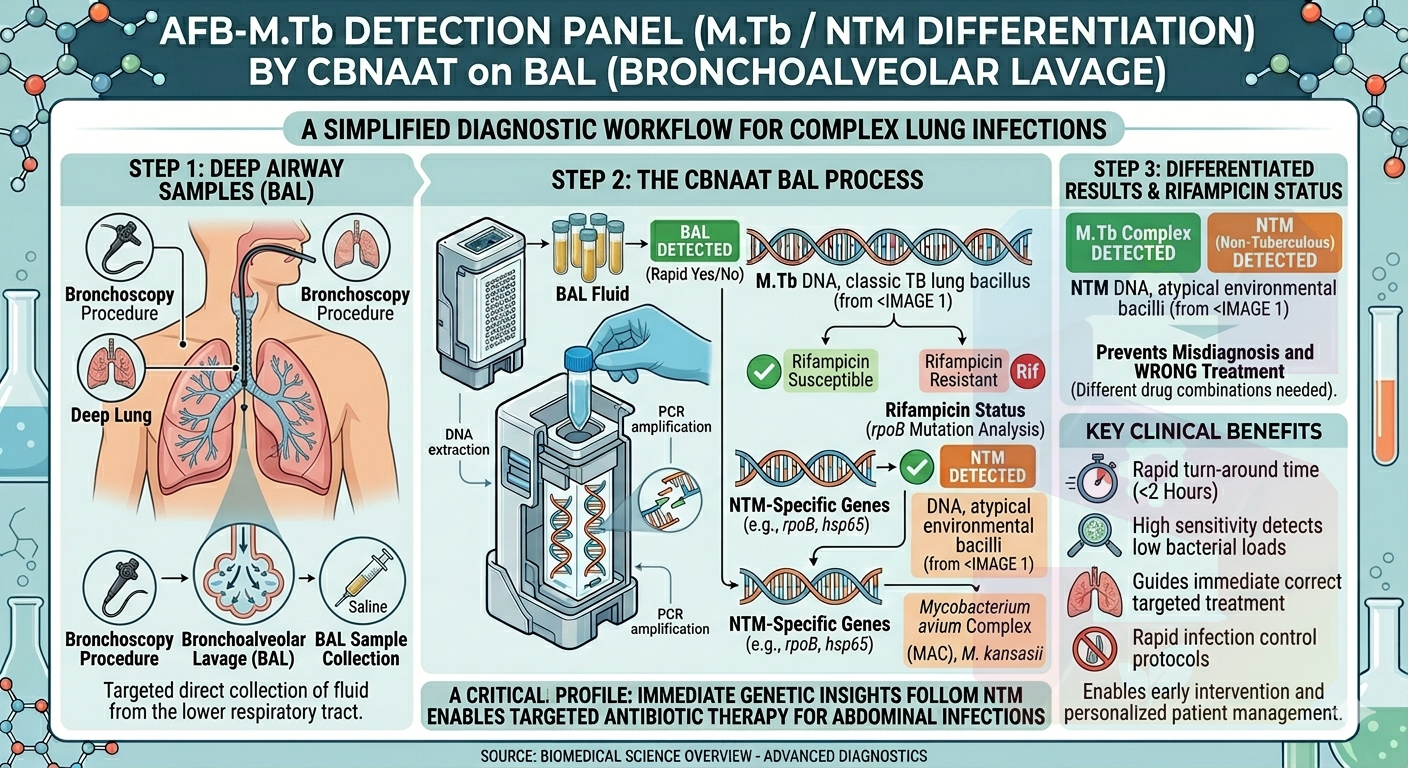
    An AFB-M.Tb Detection (M.Tb/NTM Detection) by CBNAAT on BAL (Bronchoalveolar Lavage) fluid is an advanced, rapid molecular test used to diagnose complex lung infections.

    This test simultaneously looks for classic Tuberculosis (M.Tb) and Non-Tuberculous Mycobacteria (NTM). It is specifically performed on fluid collected directly from the deep airways of the lungs during a bronchoscopy procedure.

    1. Understanding the Terms
    BAL (Bronchoalveolar Lavage): A procedure where a small scope is passed into the lungs and a sterile saline solution is used to flush out and collect cells and secretions from the deep airways. This is often done when a patient cannot produce regular sputum or when previous sputum tests were inconclusive.

    M.Tb: Mycobacterium tuberculosis, the bacteria responsible for classic, contagious pulmonary TB.

    NTM: Non-Tuberculous Mycobacteria. These are environmental bacteria that can cause lung disease mimicking TB, but they are generally non-contagious and require a completely different combination of antibiotics.

    CBNAAT: Cartridge-Based Nucleic Acid Amplification Test. An automated PCR system that multiplies and detects the unique DNA sequences of these bacteria within 2 hours.

    2. Why BAL Fluid is Used
    BAL fluid is highly valuable for diagnosing tough respiratory cases:

    Deep Lung Access: It captures samples directly from the lower respiratory tract, where the infection is actively residing.

    Overcoming Low Bacterial Counts: Traditional microscope smears require a high concentration of bacteria to show a positive result. Because CBNAAT is highly sensitive and multiplies DNA, running it on BAL fluid catches early-stage or hidden infections that standard sputum tests miss.
    An AFB-M.Tb Detection (M.Tb/NTM Detection) by CBNAAT on BAL (Bronchoalveolar Lavage) fluid is an advanced, rapid molecular test used to diagnose complex lung infections. This test simultaneously looks for classic Tuberculosis (M.Tb) and Non-Tuberculous Mycobacteria (NTM). It is specifically performed on fluid collected directly from the deep airways of the lungs during a bronchoscopy procedure. 1. Understanding the Terms BAL (Bronchoalveolar Lavage): A procedure where a small scope is passed into the lungs and a sterile saline solution is used to flush out and collect cells and secretions from the deep airways. This is often done when a patient cannot produce regular sputum or when previous sputum tests were inconclusive. M.Tb: Mycobacterium tuberculosis, the bacteria responsible for classic, contagious pulmonary TB. NTM: Non-Tuberculous Mycobacteria. These are environmental bacteria that can cause lung disease mimicking TB, but they are generally non-contagious and require a completely different combination of antibiotics. CBNAAT: Cartridge-Based Nucleic Acid Amplification Test. An automated PCR system that multiplies and detects the unique DNA sequences of these bacteria within 2 hours. 2. Why BAL Fluid is Used BAL fluid is highly valuable for diagnosing tough respiratory cases: Deep Lung Access: It captures samples directly from the lower respiratory tract, where the infection is actively residing. Overcoming Low Bacterial Counts: Traditional microscope smears require a high concentration of bacteria to show a positive result. Because CBNAAT is highly sensitive and multiplies DNA, running it on BAL fluid catches early-stage or hidden infections that standard sputum tests miss.
    0 Comments 0 Shares 619 Views 0 Reviews
  • AFB-Xpert Panel (M.Tb Detection & Rifamipicin resistance) by CBNAAT - pulmonary samples New
    2,750.00₹
    In stock
    Hyderabad
    0 Reviews
    An AFB-Xpert Panel (M.Tb Detection & Rifampicin Resistance) by CBNAAT for pulmonary samples is a highly efficient, automated molecular test. It serves as the frontline tool for rapidly diagnosing Tuberculosis (TB) in the lungs and checking if the bacteria are resistant to one of the most powerful first-line TB medications, Rifampicin.

    Unlike extra-pulmonary testing, which deals with fluids and tissues from other organs, pulmonary testing specifically evaluates secretions from the respiratory tract.

    1. What does the name mean?
    AFB: Acid-Fast Bacilli (the class of bacteria to which Mycobacterium tuberculosis belongs).

    CBNAAT: Cartridge-Based Nucleic Acid Amplification Test (commonly known as the GeneXpert test). It uses PCR technology to multiply and detect the DNA of the TB bacteria.

    Pulmonary samples: Samples originating from the lungs and respiratory airways.

    2. Common Pulmonary Sample Types
    Because pulmonary TB directly affects the lungs, the sample types are respiratory secretions:

    Sputum (Spontaneous): The thick mucus coughed up deeply from the lungs. This is the most common sample type.

    Induced Sputum: If a patient cannot cough up sputum naturally, they inhale a sterile saline mist to help loosen secretions.

    Bronchoalveolar Lavage (BAL): Fluid collected during a bronchoscopy, where a doctor passes a small scope into the lungs and flushes the area with saline. This is often used for complex or hard-to-diagnose cases.

    Gastric Aspirate/Lavage: Commonly used in young children who cannot expectorate (spit out) sputum and instead swallow their respiratory secretions overnight.
    An AFB-Xpert Panel (M.Tb Detection & Rifampicin Resistance) by CBNAAT for pulmonary samples is a highly efficient, automated molecular test. It serves as the frontline tool for rapidly diagnosing Tuberculosis (TB) in the lungs and checking if the bacteria are resistant to one of the most powerful first-line TB medications, Rifampicin. Unlike extra-pulmonary testing, which deals with fluids and tissues from other organs, pulmonary testing specifically evaluates secretions from the respiratory tract. 1. What does the name mean? AFB: Acid-Fast Bacilli (the class of bacteria to which Mycobacterium tuberculosis belongs). CBNAAT: Cartridge-Based Nucleic Acid Amplification Test (commonly known as the GeneXpert test). It uses PCR technology to multiply and detect the DNA of the TB bacteria. Pulmonary samples: Samples originating from the lungs and respiratory airways. 2. Common Pulmonary Sample Types Because pulmonary TB directly affects the lungs, the sample types are respiratory secretions: Sputum (Spontaneous): The thick mucus coughed up deeply from the lungs. This is the most common sample type. Induced Sputum: If a patient cannot cough up sputum naturally, they inhale a sterile saline mist to help loosen secretions. Bronchoalveolar Lavage (BAL): Fluid collected during a bronchoscopy, where a doctor passes a small scope into the lungs and flushes the area with saline. This is often used for complex or hard-to-diagnose cases. Gastric Aspirate/Lavage: Commonly used in young children who cannot expectorate (spit out) sputum and instead swallow their respiratory secretions overnight.
    0 Comments 0 Shares 723 Views 0 Reviews
  • AFB - detection of DNA by Real time PCR tissue and Biopsy New
    2,700.00₹
    In stock
    Hyderabad
    0 Reviews
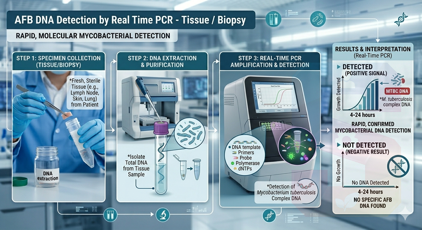
    An **AFB (Acid-Fast Bacilli) Detection of DNA by Real-Time PCR (Tissue/Biopsy)** is a highly precise molecular diagnostic test used to rapidly identify *Mycobacterium tuberculosis* complex (MTBC) and other mycobacteria directly from tissue specimens or biopsy samples.

    Unlike traditional cultures that require weeks for bacteria to grow, this test looks for the genetic blueprint (DNA) of the bacteria, offering highly accurate results in just a matter of hours.

    ---

    ### How the Test Works

    * **The Target:** The test utilizes **Real-Time Polymerase Chain Reaction (PCR)** technology to detect specific, highly conserved DNA sequences unique to mycobacteria (such as the *IS6110* or *senX3-regX3* genetic markers for *M. tuberculosis*).
    * **Amplification:** The laboratory extracts DNA from the biopsy sample (e.g., lymph node, skin, or bone tissue). If even a tiny fragment of target bacterial DNA is present, the PCR machine replicates it millions of times.
    * **Real-Time Monitoring:** Fluorescent dyes attach to the DNA copies as they are being made. A computer monitors this fluorescence in real-time; if the signal crosses a specific threshold, the test is confirmed positive.

    ---

    ### Key Advantages of This Test

    * **Exceptional Speed:** While an automated MGIT liquid culture takes 1 to 3 weeks to flag a positive result, Real-Time PCR can deliver definitive answers within **24 to 48 hours**.
    * **High Sensitivity:** It can detect minute amounts of bacterial DNA, making it incredibly useful for "paucibacillary" samples—tissue biopsies where the number of actual bacterial cells is very low.
    * **Detects Dead Bacteria:** Because it looks for DNA rather than live organisms, it can still detect the infection even if the patient has already started taking anti-TB medications.
    * **Drug Resistance Detection (Reflex):** Many modern Real-Time PCR assays (like GeneXpert or custom multiplex panels) don't just detect the DNA—they simultaneously check for mutations in genes like *rpoB*, immediately alerting doctors if the strain is resistant to major frontline antibiotics like Rifampicin.
    An **AFB (Acid-Fast Bacilli) Detection of DNA by Real-Time PCR (Tissue/Biopsy)** is a highly precise molecular diagnostic test used to rapidly identify *Mycobacterium tuberculosis* complex (MTBC) and other mycobacteria directly from tissue specimens or biopsy samples. Unlike traditional cultures that require weeks for bacteria to grow, this test looks for the genetic blueprint (DNA) of the bacteria, offering highly accurate results in just a matter of hours. --- ### How the Test Works * **The Target:** The test utilizes **Real-Time Polymerase Chain Reaction (PCR)** technology to detect specific, highly conserved DNA sequences unique to mycobacteria (such as the *IS6110* or *senX3-regX3* genetic markers for *M. tuberculosis*). * **Amplification:** The laboratory extracts DNA from the biopsy sample (e.g., lymph node, skin, or bone tissue). If even a tiny fragment of target bacterial DNA is present, the PCR machine replicates it millions of times. * **Real-Time Monitoring:** Fluorescent dyes attach to the DNA copies as they are being made. A computer monitors this fluorescence in real-time; if the signal crosses a specific threshold, the test is confirmed positive. --- ### Key Advantages of This Test * **Exceptional Speed:** While an automated MGIT liquid culture takes 1 to 3 weeks to flag a positive result, Real-Time PCR can deliver definitive answers within **24 to 48 hours**. * **High Sensitivity:** It can detect minute amounts of bacterial DNA, making it incredibly useful for "paucibacillary" samples—tissue biopsies where the number of actual bacterial cells is very low. * **Detects Dead Bacteria:** Because it looks for DNA rather than live organisms, it can still detect the infection even if the patient has already started taking anti-TB medications. * **Drug Resistance Detection (Reflex):** Many modern Real-Time PCR assays (like GeneXpert or custom multiplex panels) don't just detect the DNA—they simultaneously check for mutations in genes like *rpoB*, immediately alerting doctors if the strain is resistant to major frontline antibiotics like Rifampicin.
    0 Comments 0 Shares 830 Views 0 Reviews
  • SAT exam date in India 2026The SAT exam in India is conducted several times a year, giving students multiple opportunities to take the test based on their preparation and university deadlines. https://myflyboard.com/blog/sat-exam-dates-in-india-2026-complete-schedule-for-you
    SAT exam date in India 2026The SAT exam in India is conducted several times a year, giving students multiple opportunities to take the test based on their preparation and university deadlines. https://myflyboard.com/blog/sat-exam-dates-in-india-2026-complete-schedule-for-you
    MYFLYBOARD.COM
    SAT Exam Dates in India 2026
    Check the complete SAT Exam Dates 2026 in India. Stay updated with the full schedule, registration deadlines, and important test day details for SAT 2026.
    0 Comments 0 Shares 299 Views 0 Reviews
  • 24 Hours Urinary Protein New
    450.00₹
    In stock
    0 Reviews
    The 24-Hour Urinary Protein test is a quantitative measure of the total amount of protein excreted in the urine over a full 24-hour period. It's an important tool for diagnosing and monitoring kidney health.

    🔎 Purpose and Principle
    Purpose: To detect and quantify proteinuria (excess protein in the urine). The kidneys normally filter blood, reabsorbing almost all protein; large amounts of protein in the urine suggest kidney damage or disease.

    Principle: The test requires collecting all urine produced over 24 hours to account for the natural daily fluctuations in protein excretion. This provides the most accurate measure of total daily protein loss.
    The 24-Hour Urinary Protein test is a quantitative measure of the total amount of protein excreted in the urine over a full 24-hour period. It's an important tool for diagnosing and monitoring kidney health. 🔎 Purpose and Principle Purpose: To detect and quantify proteinuria (excess protein in the urine). The kidneys normally filter blood, reabsorbing almost all protein; large amounts of protein in the urine suggest kidney damage or disease. Principle: The test requires collecting all urine produced over 24 hours to account for the natural daily fluctuations in protein excretion. This provides the most accurate measure of total daily protein loss.
    0 Comments 0 Shares 1K Views 0 Reviews
  • 5q Deletion/Monosomy (MDS), Heparin Bone Marrow New
    5,195.00₹
    In stock
    0 Reviews
    5q Deletion/Monosomy (MDS)This refers to the 5q- syndrome, a specific subtype of Myelodysplastic Syndrome (MDS), which is a clonal hematopoietic stem cell disorder.

    Genetic Abnormality: Characterized by a deletion of part of the long arm (q-arm)3 of chromosome 5, typically del(5q), as the sole cytogenetic abnormality or with one other abnormality.
    Complete loss (monosomy 5) is less common and generally carries a poorer prognosis.
    Pathogenesis: The disease results from the haploinsufficiency (loss of one copy) of multiple genes in the deleted region, particularly the Commonly Deleted Region (CDR).
    Loss of the RPS14 gene is thought to lead to macrocytic anemia and activation of the p53 tumor suppressor pathway in erythroid precursors.
    Loss of microRNAs (e.g., miR-145, miR-146a) contributes to megakaryocyte abnormalities and normal/high platelet counts.

    Clinical Features:
    Macrocytic Anemia: Often severe and transfusion-dependent.
    Platelet Count: Usually normal or elevated (thrombocytosis), which is distinct from many other forms of MDS.
    Bone Marrow: Typically hypercellular with erythroid hypoplasia (reduced red cell precursors) and characteristic abnormal megakaryocytes that are numerous, small, and have hypolobated (non-lobulated) nuclei
    Prognosis and Treatment: Considered a relatively low-risk form of MDS compared to those with complex karyotypes.
    It has a low rate of progression to Acute Myeloid Leukemia (AML).
    The drug Lenalidomide is highly effective for this subtype, often leading to transfusion independence by targeting the 5q deleted cells.
    5q Deletion/Monosomy (MDS)This refers to the 5q- syndrome, a specific subtype of Myelodysplastic Syndrome (MDS), which is a clonal hematopoietic stem cell disorder. Genetic Abnormality: Characterized by a deletion of part of the long arm (q-arm)3 of chromosome 5, typically del(5q), as the sole cytogenetic abnormality or with one other abnormality. Complete loss (monosomy 5) is less common and generally carries a poorer prognosis. Pathogenesis: The disease results from the haploinsufficiency (loss of one copy) of multiple genes in the deleted region, particularly the Commonly Deleted Region (CDR). Loss of the RPS14 gene is thought to lead to macrocytic anemia and activation of the p53 tumor suppressor pathway in erythroid precursors. Loss of microRNAs (e.g., miR-145, miR-146a) contributes to megakaryocyte abnormalities and normal/high platelet counts. Clinical Features: Macrocytic Anemia: Often severe and transfusion-dependent. Platelet Count: Usually normal or elevated (thrombocytosis), which is distinct from many other forms of MDS. Bone Marrow: Typically hypercellular with erythroid hypoplasia (reduced red cell precursors) and characteristic abnormal megakaryocytes that are numerous, small, and have hypolobated (non-lobulated) nuclei Prognosis and Treatment: Considered a relatively low-risk form of MDS compared to those with complex karyotypes. It has a low rate of progression to Acute Myeloid Leukemia (AML). The drug Lenalidomide is highly effective for this subtype, often leading to transfusion independence by targeting the 5q deleted cells.
    0 Comments 0 Shares 1K Views 0 Reviews
  • 17p Deletion p53 (MM, CLL), Heparin Blood New
    3,650.00₹
    In stock
    Hyderabad
    0 Reviews
    A genetic test (FISH test) for 17p deletion, which involves a missing piece on the p arm of chromosome 17 that includes the TP53 gene, done using a heparin blood sample in patients with Chronic Lymphocytic Leukemia (CLL) (CLL) or other blood cancers like Multiple Myeloma (MM). This test helps assess the disease's prognosis and guides treatment decisions because the 17p deletion is a high-risk factor for poor response to traditional chemotherapy and can impact long-term survival.
    A genetic test (FISH test) for 17p deletion, which involves a missing piece on the p arm of chromosome 17 that includes the TP53 gene, done using a heparin blood sample in patients with Chronic Lymphocytic Leukemia (CLL) (CLL) or other blood cancers like Multiple Myeloma (MM). This test helps assess the disease's prognosis and guides treatment decisions because the 17p deletion is a high-risk factor for poor response to traditional chemotherapy and can impact long-term survival.
    0 Comments 0 Shares 3K Views 0 Reviews
  • 13q deletion fish technique New
    5,100.00₹
    In stock
    Hyderabad
    0 Reviews
    The 13q deletion FISH (Fluorescence in situ Hybridization) technique uses fluorescently labeled DNA probes to detect specific deletion regions, typically at 13q14, on chromosome 13q. This is done by hybridizing these probes to denatured DNA from a patient's cells (often bone marrow or blood for conditions like chronic lymphocytic leukemia (CLL) or multiple myeloma) and analyzing the fluorescent signals under a microscope. The presence of one or two normal signal areas versus a lack of signal indicates a deletion, providing crucial prognostic information for certain cancers
    The 13q deletion FISH (Fluorescence in situ Hybridization) technique uses fluorescently labeled DNA probes to detect specific deletion regions, typically at 13q14, on chromosome 13q. This is done by hybridizing these probes to denatured DNA from a patient's cells (often bone marrow or blood for conditions like chronic lymphocytic leukemia (CLL) or multiple myeloma) and analyzing the fluorescent signals under a microscope. The presence of one or two normal signal areas versus a lack of signal indicates a deletion, providing crucial prognostic information for certain cancers
    0 Comments 0 Shares 3K Views 0 Reviews
Sponsored