

• Beta 2 Microglobulin test New
    2,000.00₹
    In stock
    All over India
    0 Reviews
    The beta-2 microglobulin (B2M) test measures the amount of a small protein found on the surface of most cells in your body. Small amounts naturally shed into your blood and urine, but high levels can mean certain cells are turning over too quickly or your immune system is working overtime. Doctors usually order this test to help diagnose, monitor, or check the severity of blood cancers like multiple myeloma, lymphoma, and certain leukemias. It can also help evaluate kidney damage or inflammatory conditions, giving your healthcare team important insights into your overall health and treatment progress.
    The beta-2 microglobulin (B2M) test measures the amount of a small protein found on the surface of most cells in your body. Small amounts naturally shed into your blood and urine, but high levels can mean certain cells are turning over too quickly or your immune system is working overtime. Doctors usually order this test to help diagnose, monitor, or check the severity of blood cancers like multiple myeloma, lymphoma, and certain leukemias. It can also help evaluate kidney damage or inflammatory conditions, giving your healthcare team important insights into your overall health and treatment progress.
    0 Comments 0 Shares 98 Views 0 Reviews
  • Berylium Test New
    2,145.00₹
    In stock
    All over India
    0 Reviews
    The beryllium test—most commonly performed as the Beryllium Lymphocyte Proliferation Test (BeLPT)—is a specialized immunological assay used to evaluate sensitization to the toxic metal beryllium. Exposure primarily occurs in industries such as aerospace, electronics, and metal machining. The assay measures how a patient's white blood cells react when exposed to beryllium salts in a laboratory setting. A positive proliferation response indicates cellular sensitization, which serves as a crucial diagnostic marker for chronic beryllium disease (CBD). Early detection enables clinicians to monitor lung health, manage occupational exposure risks, and implement timely interventions before irreversible pulmonary fibrosis develops.
    The beryllium test—most commonly performed as the Beryllium Lymphocyte Proliferation Test (BeLPT)—is a specialized immunological assay used to evaluate sensitization to the toxic metal beryllium. Exposure primarily occurs in industries such as aerospace, electronics, and metal machining. The assay measures how a patient's white blood cells react when exposed to beryllium salts in a laboratory setting. A positive proliferation response indicates cellular sensitization, which serves as a crucial diagnostic marker for chronic beryllium disease (CBD). Early detection enables clinicians to monitor lung health, manage occupational exposure risks, and implement timely interventions before irreversible pulmonary fibrosis develops.
    0 Comments 0 Shares 94 Views 0 Reviews
  • BCR-ABL IS(International Scale) Transcript Fusion Quantification by Real time PCR - Blood New
    6,000.00₹
    In stock
    All over India
    0 Reviews
    The BCR-ABL IS (International Scale) Transcript Fusion Quantification test is a sensitive molecular assay performed on peripheral blood using real-time reverse transcription-polymerase chain reaction (RT-PCR).
    Designed primarily for patients with chronic myeloid leukemia (CML), this test measures the quantity of specific fusion transcripts (such as p210). By standardizing the results onto the International Scale (IS), it allows clinicians to precisely track disease burden, evaluate therapeutic responses to tyrosine kinase inhibitors, and reliably monitor minimal residual disease (MRD) over time across different laboratories with high accuracy and reproducibility.
    The BCR-ABL IS (International Scale) Transcript Fusion Quantification test is a sensitive molecular assay performed on peripheral blood using real-time reverse transcription-polymerase chain reaction (RT-PCR). Designed primarily for patients with chronic myeloid leukemia (CML), this test measures the quantity of specific fusion transcripts (such as p210). By standardizing the results onto the International Scale (IS), it allows clinicians to precisely track disease burden, evaluate therapeutic responses to tyrosine kinase inhibitors, and reliably monitor minimal residual disease (MRD) over time across different laboratories with high accuracy and reproducibility.
    0 Comments 0 Shares 103 Views 0 Reviews
  • Basic Arthritis Profile for Male New
    1,100.00₹
    In stock
    All over India
    0 Reviews
    A **Basic Arthritis Profile for Male** is a targeted blood panel designed to evaluate systemic inflammation and identify markers associated with various arthritic conditions. This profile typically includes tests such as Erythrocyte Sedimentation Rate (ESR) and C-Reactive Protein (CRP) to detect generalized inflammatory responses, along with Rheumatoid Factor (RF) and sometimes Anti-CCP to screen for autoimmune joint diseases. Additionally, Uric Acid levels are frequently measured to assess for gout, a condition notably prevalent in men. By analyzing these specific biomarkers, healthcare providers can differentiate between inflammatory and non-inflammatory joint pain, facilitating an accurate diagnosis and timely intervention to preserve long-term joint health.
    A **Basic Arthritis Profile for Male** is a targeted blood panel designed to evaluate systemic inflammation and identify markers associated with various arthritic conditions. This profile typically includes tests such as Erythrocyte Sedimentation Rate (ESR) and C-Reactive Protein (CRP) to detect generalized inflammatory responses, along with Rheumatoid Factor (RF) and sometimes Anti-CCP to screen for autoimmune joint diseases. Additionally, Uric Acid levels are frequently measured to assess for gout, a condition notably prevalent in men. By analyzing these specific biomarkers, healthcare providers can differentiate between inflammatory and non-inflammatory joint pain, facilitating an accurate diagnosis and timely intervention to preserve long-term joint health.
    0 Comments 0 Shares 305 Views 0 Reviews
  • Barium Test New
    2,145.00₹
    In stock
    All over India
    0 Reviews
    A **Barium Test**, often referred to as a barium swallow or barium meal, is a diagnostic imaging procedure used to examine the upper gastrointestinal (GI) tract. During the test, the patient consumes a liquid containing barium sulfate, a contrast medium that coats the lining of the esophagus, stomach, and small intestine. Because barium is radiopaque, it appears white on X-rays, allowing radiologists to clearly visualize the anatomy and identify abnormalities such as ulcers, tumors, strictures, or swallowing difficulties. This non-invasive assessment provides essential real-time structural and functional data, assisting clinicians in diagnosing various digestive disorders and guiding effective treatment plans.
    A **Barium Test**, often referred to as a barium swallow or barium meal, is a diagnostic imaging procedure used to examine the upper gastrointestinal (GI) tract. During the test, the patient consumes a liquid containing barium sulfate, a contrast medium that coats the lining of the esophagus, stomach, and small intestine. Because barium is radiopaque, it appears white on X-rays, allowing radiologists to clearly visualize the anatomy and identify abnormalities such as ulcers, tumors, strictures, or swallowing difficulties. This non-invasive assessment provides essential real-time structural and functional data, assisting clinicians in diagnosing various digestive disorders and guiding effective treatment plans.
    0 Comments 0 Shares 325 Views 0 Reviews
  • Back swelling- Medium Biopsy (1-3cm) New
    1,000.00₹
    In stock
    All over India
    0 Reviews
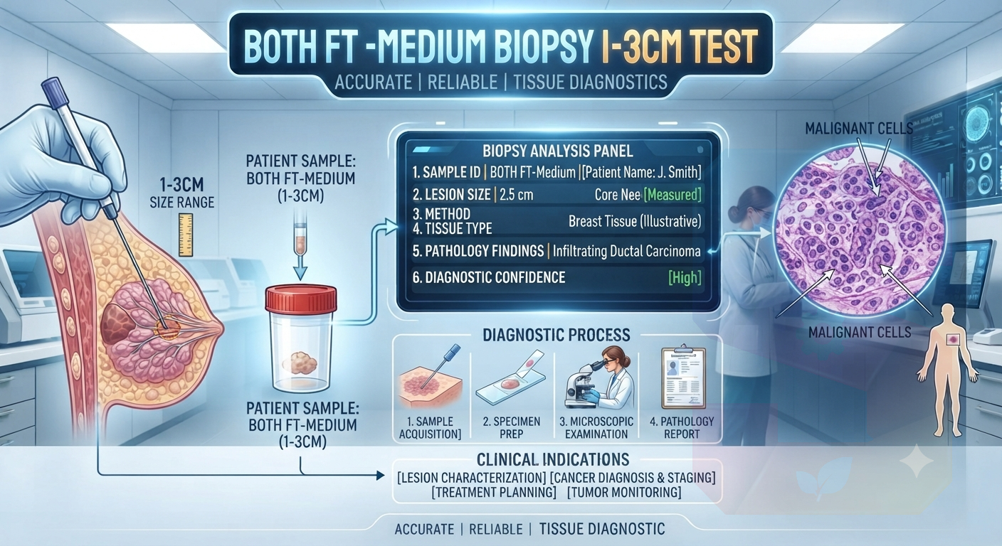
    A **Back Swelling – Medium Biopsy (1–3 cm)** refers to the surgical sampling of a subcutaneous or intramuscular growth located on the posterior torso measuring between 1 and 3 centimeters. This procedure is performed to obtain a definitive histological diagnosis, allowing clinicians to distinguish between benign entities—such as lipomas, sebaceous cysts, or fibromas—and potential malignancies. The process involves local anesthesia, an appropriately sized incision, and precise tissue extraction. Pathological evaluation of this sample is critical, as it confirms the lesion's cellular architecture and nature, directly guiding subsequent clinical management, surgical excision plans, or necessary therapeutic interventions.
    A **Back Swelling – Medium Biopsy (1–3 cm)** refers to the surgical sampling of a subcutaneous or intramuscular growth located on the posterior torso measuring between 1 and 3 centimeters. This procedure is performed to obtain a definitive histological diagnosis, allowing clinicians to distinguish between benign entities—such as lipomas, sebaceous cysts, or fibromas—and potential malignancies. The process involves local anesthesia, an appropriately sized incision, and precise tissue extraction. Pathological evaluation of this sample is critical, as it confirms the lesion's cellular architecture and nature, directly guiding subsequent clinical management, surgical excision plans, or necessary therapeutic interventions.
    0 Comments 0 Shares 305 Views 0 Reviews
  • Back swelling- large Biopsy (3-6cm) New
    1,500.00₹
    In stock
    All over India
    0 Reviews
    Back Swelling – Large Biopsy (3–6 cm)

    A Back Swelling – Large Biopsy (3–6 cm) is a procedure in which a tissue sample is taken from a larger lump or swelling on the back for laboratory examination. It helps identify the exact cause, such as a benign growth, infection, inflammation, cyst, or cancer. The biopsy is usually performed under local anesthesia, making it a safe and minimally uncomfortable procedure. Accurate diagnosis allows doctors to plan the most appropriate treatment and avoid unnecessary surgery. If you notice a persistent, enlarging, or painful back swelling, consult your healthcare provider promptly for proper evaluation and timely management.
    Back Swelling – Large Biopsy (3–6 cm) A Back Swelling – Large Biopsy (3–6 cm) is a procedure in which a tissue sample is taken from a larger lump or swelling on the back for laboratory examination. It helps identify the exact cause, such as a benign growth, infection, inflammation, cyst, or cancer. The biopsy is usually performed under local anesthesia, making it a safe and minimally uncomfortable procedure. Accurate diagnosis allows doctors to plan the most appropriate treatment and avoid unnecessary surgery. If you notice a persistent, enlarging, or painful back swelling, consult your healthcare provider promptly for proper evaluation and timely management.
    0 Comments 0 Shares 248 Views 0 Reviews
  • Autoimmune Liver disease Profile - 1 New
    4,500.00₹
    In stock
    All over India
    0 Reviews
    The **Autoimmune Liver Disease Profile 1** is a specialized diagnostic test package designed to identify autoimmune etiologies of liver inflammation. This panel typically screens for a combination of highly specific autoantibodies—such as Anti-Nuclear Antibodies (ANA), Anti-Smooth Muscle Antibodies (ASMA), and Anti-Liver-Kidney Microsomal (LKM) antibodies—which are clinical markers for conditions like Autoimmune Hepatitis, Primary Biliary Cholangitis, or Primary Sclerosing Cholangitis. By detecting these characteristic immune signatures, the profile enables clinicians to accurately differentiate autoimmune liver disorders from other hepatic pathologies. Early identification through this comprehensive antibody assessment allows for the timely initiation of targeted immunosuppressive therapy to preserve liver function and prevent progression to cirrhosis.
    The **Autoimmune Liver Disease Profile 1** is a specialized diagnostic test package designed to identify autoimmune etiologies of liver inflammation. This panel typically screens for a combination of highly specific autoantibodies—such as Anti-Nuclear Antibodies (ANA), Anti-Smooth Muscle Antibodies (ASMA), and Anti-Liver-Kidney Microsomal (LKM) antibodies—which are clinical markers for conditions like Autoimmune Hepatitis, Primary Biliary Cholangitis, or Primary Sclerosing Cholangitis. By detecting these characteristic immune signatures, the profile enables clinicians to accurately differentiate autoimmune liver disorders from other hepatic pathologies. Early identification through this comprehensive antibody assessment allows for the timely initiation of targeted immunosuppressive therapy to preserve liver function and prevent progression to cirrhosis.
    0 Comments 0 Shares 497 Views 0 Reviews
  • Arthritis Screen Profile (4 marker) New
    1,081.00₹
    In stock
    All over India
    0 Reviews
    An **Arthritis Screen Profile (4 Markers)** is a targeted, focused diagnostic blood test designed for early evaluation of joint pain and systemic inflammation. By measuring four key biomarkers—typically **Rheumatoid Factor (RF)**, **Anti-CCP**, **C-Reactive Protein (CRP)**, and **Erythrocyte Sedimentation Rate (ESR)**—this profile helps clinicians differentiate between autoimmune disorders like rheumatoid arthritis and non-autoimmune conditions.
    The test detects active inflammatory processes, identifies specific autoantibodies, and assesses overall disease severity. Swift and precise, the 4-marker panel enables healthcare providers to establish an accurate diagnosis, start timely therapeutic intervention, and help safeguard long-term joint health and mobility.
    An **Arthritis Screen Profile (4 Markers)** is a targeted, focused diagnostic blood test designed for early evaluation of joint pain and systemic inflammation. By measuring four key biomarkers—typically **Rheumatoid Factor (RF)**, **Anti-CCP**, **C-Reactive Protein (CRP)**, and **Erythrocyte Sedimentation Rate (ESR)**—this profile helps clinicians differentiate between autoimmune disorders like rheumatoid arthritis and non-autoimmune conditions. The test detects active inflammatory processes, identifies specific autoantibodies, and assesses overall disease severity. Swift and precise, the 4-marker panel enables healthcare providers to establish an accurate diagnosis, start timely therapeutic intervention, and help safeguard long-term joint health and mobility.
    0 Comments 0 Shares 319 Views 0 Reviews
  • Arthritis Scan Test New
    1,099.00₹
    In stock
    All over India
    0 Reviews
    An **arthritis scan test** is a non-invasive diagnostic imaging procedure—such as an MRI, ultrasound, X-ray, or CT scan—used to evaluate joint health and detect structural changes. It provides detailed visual insights into affected joints, allowing clinicians to assess cartilage wear, bone erosion, joint space narrowing, fluid accumulation, and soft tissue inflammation.
    By accurately identifying conditions like osteoarthritis, rheumatoid arthritis, or psoriatic arthritis, these scans play a critical role in confirming diagnoses. They also help monitor disease progression and evaluate treatment response over time. Timely imaging empowers healthcare providers to customize targeted therapies, protect joint functionality, and manage long-term discomfort effectively.
    An **arthritis scan test** is a non-invasive diagnostic imaging procedure—such as an MRI, ultrasound, X-ray, or CT scan—used to evaluate joint health and detect structural changes. It provides detailed visual insights into affected joints, allowing clinicians to assess cartilage wear, bone erosion, joint space narrowing, fluid accumulation, and soft tissue inflammation. By accurately identifying conditions like osteoarthritis, rheumatoid arthritis, or psoriatic arthritis, these scans play a critical role in confirming diagnoses. They also help monitor disease progression and evaluate treatment response over time. Timely imaging empowers healthcare providers to customize targeted therapies, protect joint functionality, and manage long-term discomfort effectively.
    0 Comments 0 Shares 319 Views 0 Reviews
  • APTT New
    418.00₹
    In stock
    All over India
    0 Reviews
    The **Activated Partial Thromboplastin Time (APTT)** test is a essential coagulation screening assay that evaluates the integrity of the intrinsic and common pathways of the blood clotting cascade. It measures the time, in seconds, required for a fibrin clot to form in plasma after specific activating reagents and calcium are added. Clinically, the APTT is primary used to monitor unfractionated heparin anticoagulant therapy and to investigate unexplained bleeding or bruising. Prolonged APTT values can indicate deficiency or dysfunction of clotting factors—such as Factor VIII in Hemophilia A or Factor IX in Hemophilia B—or the presence of circulating inhibitors like lupus anticoagulant.
    The **Activated Partial Thromboplastin Time (APTT)** test is a essential coagulation screening assay that evaluates the integrity of the intrinsic and common pathways of the blood clotting cascade. It measures the time, in seconds, required for a fibrin clot to form in plasma after specific activating reagents and calcium are added. Clinically, the APTT is primary used to monitor unfractionated heparin anticoagulant therapy and to investigate unexplained bleeding or bruising. Prolonged APTT values can indicate deficiency or dysfunction of clotting factors—such as Factor VIII in Hemophilia A or Factor IX in Hemophilia B—or the presence of circulating inhibitors like lupus anticoagulant.
    0 Comments 0 Shares 1K Views 0 Reviews
  • Appendix medium biopsy 1 -3cm New
    1,000.00₹
    In stock
    All over India
    0 Reviews
    The **Appendix medium Biopsy 1-3cm Test** is a highly specific pathological examination performed on a surgically removed appendix specimen that presents an abnormal lesion or segment within the 1 to 3-centimeter size range. Unlike standard appendectomy reviews, this test requires critical microscopic analysis of the entire specimen, as well as multiple distinct tissue slices, to evaluate architectural complexity and check for pre-malignant changes. Pathologists meticulously look for early-stage neuroendocrine tumors, mucinous neoplasms, or sessile serrated lesions that may be hidden in this size category. The examination results provide vital diagnostic clarity, distinguishing between benign inflammation and subtle cancers, which directly guides subsequent patient management, such as the potential need for right hemicolectomy or long-term surveillance.
    The **Appendix medium Biopsy 1-3cm Test** is a highly specific pathological examination performed on a surgically removed appendix specimen that presents an abnormal lesion or segment within the 1 to 3-centimeter size range. Unlike standard appendectomy reviews, this test requires critical microscopic analysis of the entire specimen, as well as multiple distinct tissue slices, to evaluate architectural complexity and check for pre-malignant changes. Pathologists meticulously look for early-stage neuroendocrine tumors, mucinous neoplasms, or sessile serrated lesions that may be hidden in this size category. The examination results provide vital diagnostic clarity, distinguishing between benign inflammation and subtle cancers, which directly guides subsequent patient management, such as the potential need for right hemicolectomy or long-term surveillance.
    0 Comments 0 Shares 1K Views 0 Reviews
More Results
Sponsored