

• Beta 2 Glycoprotein 1 - IgA Test New
    1,500.00₹
    In stock
    All over India
    0 Reviews
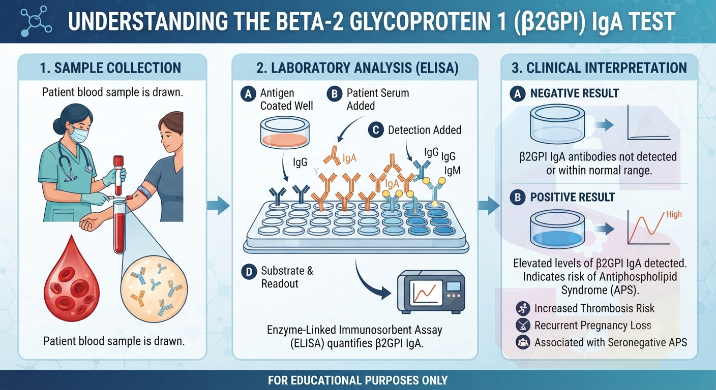
    The Beta-2 Glycoprotein 1 (β2GPI) IgA test is a specialized immunoassay used to detect IgA-class autoantibodies directed against β2GPI, a plasma protein involved in blood coagulation. While IgG and IgM are the primary isotypes associated with Antiphospholipid Syndrome (APS), measuring IgA provides critical diagnostic value. It is particularly useful in evaluating patients with strong clinical suspicion of APS—such as unexplained thrombosis or recurrent pregnancy loss—who test negative for conventional criteria antibodies. Detecting isolated β2GPI IgA antibodies aids in the comprehensive risk stratification and diagnosis of seronegative APS, guiding appropriate long-term management and thromboprophylaxis.
    The Beta-2 Glycoprotein 1 (β2GPI) IgA test is a specialized immunoassay used to detect IgA-class autoantibodies directed against β2GPI, a plasma protein involved in blood coagulation. While IgG and IgM are the primary isotypes associated with Antiphospholipid Syndrome (APS), measuring IgA provides critical diagnostic value. It is particularly useful in evaluating patients with strong clinical suspicion of APS—such as unexplained thrombosis or recurrent pregnancy loss—who test negative for conventional criteria antibodies. Detecting isolated β2GPI IgA antibodies aids in the comprehensive risk stratification and diagnosis of seronegative APS, guiding appropriate long-term management and thromboprophylaxis.
    0 Comments 0 Shares 317 Views 0 Reviews
  • Bence Jones Protein -Qualitative New
    1,350.00₹
    In stock
    All over India
    0 Reviews
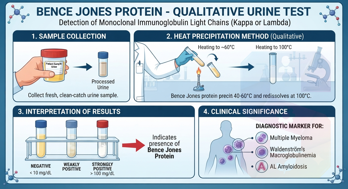
    The Bence Jones Protein qualitative test is a laboratory analysis used to detect free monoclonal immunoglobulin light chains (kappa or lambda) in urine. These abnormal proteins are frequently produced in excess by plasma cell disorders, most notably multiple myeloma, as well as Waldenström macroglobulinemia and amyloidosis. Detecting Bence Jones proteins aids clinicians in diagnosing and monitoring these conditions. While a qualitative test provides a straightforward positive or negative result to indicate their presence, it serves as a critical initial screening marker that often prompts further quantitative evaluation and comprehensive clinical workups for underlying monoclonal gammopathies.
    The Bence Jones Protein qualitative test is a laboratory analysis used to detect free monoclonal immunoglobulin light chains (kappa or lambda) in urine. These abnormal proteins are frequently produced in excess by plasma cell disorders, most notably multiple myeloma, as well as Waldenström macroglobulinemia and amyloidosis. Detecting Bence Jones proteins aids clinicians in diagnosing and monitoring these conditions. While a qualitative test provides a straightforward positive or negative result to indicate their presence, it serves as a critical initial screening marker that often prompts further quantitative evaluation and comprehensive clinical workups for underlying monoclonal gammopathies.
    0 Comments 0 Shares 314 Views 0 Reviews
  • Barium Test New
    2,145.00₹
    In stock
    All over India
    0 Reviews
    A **Barium Test**, often referred to as a barium swallow or barium meal, is a diagnostic imaging procedure used to examine the upper gastrointestinal (GI) tract. During the test, the patient consumes a liquid containing barium sulfate, a contrast medium that coats the lining of the esophagus, stomach, and small intestine. Because barium is radiopaque, it appears white on X-rays, allowing radiologists to clearly visualize the anatomy and identify abnormalities such as ulcers, tumors, strictures, or swallowing difficulties. This non-invasive assessment provides essential real-time structural and functional data, assisting clinicians in diagnosing various digestive disorders and guiding effective treatment plans.
    A **Barium Test**, often referred to as a barium swallow or barium meal, is a diagnostic imaging procedure used to examine the upper gastrointestinal (GI) tract. During the test, the patient consumes a liquid containing barium sulfate, a contrast medium that coats the lining of the esophagus, stomach, and small intestine. Because barium is radiopaque, it appears white on X-rays, allowing radiologists to clearly visualize the anatomy and identify abnormalities such as ulcers, tumors, strictures, or swallowing difficulties. This non-invasive assessment provides essential real-time structural and functional data, assisting clinicians in diagnosing various digestive disorders and guiding effective treatment plans.
    0 Comments 0 Shares 430 Views 0 Reviews
  • Back swelling- Medium Biopsy (1-3cm) New
    1,000.00₹
    In stock
    All over India
    0 Reviews
    A **Back Swelling – Medium Biopsy (1–3 cm)** refers to the surgical sampling of a subcutaneous or intramuscular growth located on the posterior torso measuring between 1 and 3 centimeters. This procedure is performed to obtain a definitive histological diagnosis, allowing clinicians to distinguish between benign entities—such as lipomas, sebaceous cysts, or fibromas—and potential malignancies. The process involves local anesthesia, an appropriately sized incision, and precise tissue extraction. Pathological evaluation of this sample is critical, as it confirms the lesion's cellular architecture and nature, directly guiding subsequent clinical management, surgical excision plans, or necessary therapeutic interventions.
    A **Back Swelling – Medium Biopsy (1–3 cm)** refers to the surgical sampling of a subcutaneous or intramuscular growth located on the posterior torso measuring between 1 and 3 centimeters. This procedure is performed to obtain a definitive histological diagnosis, allowing clinicians to distinguish between benign entities—such as lipomas, sebaceous cysts, or fibromas—and potential malignancies. The process involves local anesthesia, an appropriately sized incision, and precise tissue extraction. Pathological evaluation of this sample is critical, as it confirms the lesion's cellular architecture and nature, directly guiding subsequent clinical management, surgical excision plans, or necessary therapeutic interventions.
    0 Comments 0 Shares 327 Views 0 Reviews
  • Back swelling- large Biopsy (3-6cm) New
    1,500.00₹
    In stock
    All over India
    0 Reviews
    Back Swelling – Large Biopsy (3–6 cm)

    A Back Swelling – Large Biopsy (3–6 cm) is a procedure in which a tissue sample is taken from a larger lump or swelling on the back for laboratory examination. It helps identify the exact cause, such as a benign growth, infection, inflammation, cyst, or cancer. The biopsy is usually performed under local anesthesia, making it a safe and minimally uncomfortable procedure. Accurate diagnosis allows doctors to plan the most appropriate treatment and avoid unnecessary surgery. If you notice a persistent, enlarging, or painful back swelling, consult your healthcare provider promptly for proper evaluation and timely management.
    Back Swelling – Large Biopsy (3–6 cm) A Back Swelling – Large Biopsy (3–6 cm) is a procedure in which a tissue sample is taken from a larger lump or swelling on the back for laboratory examination. It helps identify the exact cause, such as a benign growth, infection, inflammation, cyst, or cancer. The biopsy is usually performed under local anesthesia, making it a safe and minimally uncomfortable procedure. Accurate diagnosis allows doctors to plan the most appropriate treatment and avoid unnecessary surgery. If you notice a persistent, enlarging, or painful back swelling, consult your healthcare provider promptly for proper evaluation and timely management.
    0 Comments 0 Shares 278 Views 0 Reviews
  • Arthritis Profile - B Test New
    4,000.00₹
    In stock
    All over India
    0 Reviews
    An **Arthritis Profile B test** is an expanded, multi-parameter diagnostic panel designed to comprehensively evaluate complex joint disorders and autoimmune inflammatory conditions. In addition to core markers like **Rheumatoid Factor (RF)**, **C-Reactive Protein (CRP)**, and **Erythrocyte Sedimentation Rate (ESR)**, Profile B typically incorporates specialized biomarkers such as **Anti-CCP**, **Anti-Nuclear Antibodies (ANA)**, **ASO Titre**, **HLA-B27**, **Complement 3 (C3)**, and **Vitamin D levels**.
    Clinicians order this extensive profile for patients presenting with persistent joint pain, morning stiffness, or suspected systemic autoimmune diseases like ankylosing spondylitis or lupus. It provides deep diagnostic insight to differentiate overlapping arthritic conditions and guide targeted therapy.
    An **Arthritis Profile B test** is an expanded, multi-parameter diagnostic panel designed to comprehensively evaluate complex joint disorders and autoimmune inflammatory conditions. In addition to core markers like **Rheumatoid Factor (RF)**, **C-Reactive Protein (CRP)**, and **Erythrocyte Sedimentation Rate (ESR)**, Profile B typically incorporates specialized biomarkers such as **Anti-CCP**, **Anti-Nuclear Antibodies (ANA)**, **ASO Titre**, **HLA-B27**, **Complement 3 (C3)**, and **Vitamin D levels**. Clinicians order this extensive profile for patients presenting with persistent joint pain, morning stiffness, or suspected systemic autoimmune diseases like ankylosing spondylitis or lupus. It provides deep diagnostic insight to differentiate overlapping arthritic conditions and guide targeted therapy.
    0 Comments 0 Shares 458 Views 0 Reviews
  • Arthritis Basic Screening Test New
    1,499.00₹
    In stock
    All over India
    0 Reviews
    An **Arthritis Basic Screening Test** is a panel of routine blood tests designed to detect inflammation, evaluate joint health, and identify underlying causes of joint pain and stiffness. The panel typically measures key inflammatory markers, such as **C-Reactive Protein (CRP)** and **Erythrocyte Sedimentation Rate (ESR)**, along with a **Rheumatoid Factor (RF)** test to help differentiate rheumatoid arthritis from other joint disorders. It often includes a **Uric Acid test** to evaluate for gout.
    Clinicians use this basic profile as an initial diagnostic step to guide appropriate treatment, monitor disease progression, and refer patients to a rheumatologist.
    An **Arthritis Basic Screening Test** is a panel of routine blood tests designed to detect inflammation, evaluate joint health, and identify underlying causes of joint pain and stiffness. The panel typically measures key inflammatory markers, such as **C-Reactive Protein (CRP)** and **Erythrocyte Sedimentation Rate (ESR)**, along with a **Rheumatoid Factor (RF)** test to help differentiate rheumatoid arthritis from other joint disorders. It often includes a **Uric Acid test** to evaluate for gout. Clinicians use this basic profile as an initial diagnostic step to guide appropriate treatment, monitor disease progression, and refer patients to a rheumatologist.
    0 Comments 0 Shares 485 Views 0 Reviews
  • Arm swelling biopsy - medium 1-3cm New
    1,000.00₹
    In stock
    All over India
    0 Reviews
    An arm swelling biopsy (medium 1-3 cm) is a medical procedure used to obtain a tissue sample from an abnormal growth in the arm. Typically, this applies to palpable masses, deep-seated lesions, or lymph nodes within that size range. A dermatologist or surgeon performs either an incisional biopsy to remove a portion or an excisional biopsy to remove the entire mass. The procedure is usually ultrasound-guided to ensure precise tissue collection. The acquired specimen is crucial for a definitive pathological diagnosis, distinguishing between benign, inflammatory, or malignant conditions. The results of the biopsy are the key determinant for guiding appropriate medical management or surgical treatment.
    An arm swelling biopsy (medium 1-3 cm) is a medical procedure used to obtain a tissue sample from an abnormal growth in the arm. Typically, this applies to palpable masses, deep-seated lesions, or lymph nodes within that size range. A dermatologist or surgeon performs either an incisional biopsy to remove a portion or an excisional biopsy to remove the entire mass. The procedure is usually ultrasound-guided to ensure precise tissue collection. The acquired specimen is crucial for a definitive pathological diagnosis, distinguishing between benign, inflammatory, or malignant conditions. The results of the biopsy are the key determinant for guiding appropriate medical management or surgical treatment.
    0 Comments 0 Shares 1K Views 0 Reviews
  • Appendix medium biopsy 1 -3cm New
    1,000.00₹
    In stock
    All over India
    0 Reviews
    The **Appendix medium Biopsy 1-3cm Test** is a highly specific pathological examination performed on a surgically removed appendix specimen that presents an abnormal lesion or segment within the 1 to 3-centimeter size range. Unlike standard appendectomy reviews, this test requires critical microscopic analysis of the entire specimen, as well as multiple distinct tissue slices, to evaluate architectural complexity and check for pre-malignant changes. Pathologists meticulously look for early-stage neuroendocrine tumors, mucinous neoplasms, or sessile serrated lesions that may be hidden in this size category. The examination results provide vital diagnostic clarity, distinguishing between benign inflammation and subtle cancers, which directly guides subsequent patient management, such as the potential need for right hemicolectomy or long-term surveillance.
    The **Appendix medium Biopsy 1-3cm Test** is a highly specific pathological examination performed on a surgically removed appendix specimen that presents an abnormal lesion or segment within the 1 to 3-centimeter size range. Unlike standard appendectomy reviews, this test requires critical microscopic analysis of the entire specimen, as well as multiple distinct tissue slices, to evaluate architectural complexity and check for pre-malignant changes. Pathologists meticulously look for early-stage neuroendocrine tumors, mucinous neoplasms, or sessile serrated lesions that may be hidden in this size category. The examination results provide vital diagnostic clarity, distinguishing between benign inflammation and subtle cancers, which directly guides subsequent patient management, such as the potential need for right hemicolectomy or long-term surveillance.
    0 Comments 0 Shares 1K Views 0 Reviews
  • Appendix large biopsy 3 -6cm New
    1,500.00₹
    In stock
    All over India
    0 Reviews

    An **appendix large biopsy (3 to 6 cm)**, often performed as an excisional biopsy or an appendectomy, involves the removal of a substantial portion of the appendix for detailed pathological examination. This procedure is typically warranted when imaging or previous smaller biopsies are inconclusive, particularly to evaluate for conditions beyond acute appendicitis, such as appendiceal neoplasms, carcinoid tumors, or other complex pathological processes within the large 3 to 6 cm range. A pathologist will conduct a thorough macroscopic and microscopic assessment of the tissue structure, check surgical margins, and determine specific tumor type or grade, providing crucial data for accurate diagnosis and optimal treatment planning for complex appendiceal conditions.
    An **appendix large biopsy (3 to 6 cm)**, often performed as an excisional biopsy or an appendectomy, involves the removal of a substantial portion of the appendix for detailed pathological examination. This procedure is typically warranted when imaging or previous smaller biopsies are inconclusive, particularly to evaluate for conditions beyond acute appendicitis, such as appendiceal neoplasms, carcinoid tumors, or other complex pathological processes within the large 3 to 6 cm range. A pathologist will conduct a thorough macroscopic and microscopic assessment of the tissue structure, check surgical margins, and determine specific tumor type or grade, providing crucial data for accurate diagnosis and optimal treatment planning for complex appendiceal conditions.
    0 Comments 0 Shares 1K Views 0 Reviews
  • APLA Profile Test New
    2,999.00₹
    In stock
    All over India
    0 Reviews
    An **Antiphospholipid Antibody (APLA) Profile Test** is a specialized group of blood tests used to diagnose Antiphospholipid Syndrome (APS), an autoimmune condition. In APS, the body mistakenly produces autoantibodies against its own proteins bound to phospholipids. This reaction significantly increases the risk of abnormal blood clotting in veins or arteries, as well as pregnancy complications like recurrent miscarriages.
    The APLA panel typically measures three primary markers: lupus anticoagulant, anticardiolipin antibodies, and anti-beta-2 glycoprotein I antibodies. Identifying these autoantibodies helps clinicians evaluate a patient's risk of thrombotic events, guiding appropriate preventive care and targeted anticoagulant treatments.
    An **Antiphospholipid Antibody (APLA) Profile Test** is a specialized group of blood tests used to diagnose Antiphospholipid Syndrome (APS), an autoimmune condition. In APS, the body mistakenly produces autoantibodies against its own proteins bound to phospholipids. This reaction significantly increases the risk of abnormal blood clotting in veins or arteries, as well as pregnancy complications like recurrent miscarriages. The APLA panel typically measures three primary markers: lupus anticoagulant, anticardiolipin antibodies, and anti-beta-2 glycoprotein I antibodies. Identifying these autoantibodies helps clinicians evaluate a patient's risk of thrombotic events, guiding appropriate preventive care and targeted anticoagulant treatments.
    0 Comments 0 Shares 992 Views 0 Reviews
  • Any lumps and Bumps New
    1,000.00₹
    In stock
    All over India
    0 Reviews
    An **Any Lumps and Bumps Test** evaluation refers to a clinical diagnostic approach used to investigate unexplained swellings, masses, or cutaneous nodules on the body.
    Rather than a single blood test, this assessment typically combines physical examination with imaging modalities like **ultrasound**, **MRI**, or a **fine-needle aspiration (FNA) biopsy** to determine the characteristics of the lump.
    The primary utility is differentiating between benign conditions (such as lipomas, sebaceous cysts, or swollen lymph nodes) and malignant tumors. Identifying the exact nature of the tissue growth ensures accurate staging, patient reassurance, and the timely implementation of appropriate medical or surgical intervention strategies.
    An **Any Lumps and Bumps Test** evaluation refers to a clinical diagnostic approach used to investigate unexplained swellings, masses, or cutaneous nodules on the body. Rather than a single blood test, this assessment typically combines physical examination with imaging modalities like **ultrasound**, **MRI**, or a **fine-needle aspiration (FNA) biopsy** to determine the characteristics of the lump. The primary utility is differentiating between benign conditions (such as lipomas, sebaceous cysts, or swollen lymph nodes) and malignant tumors. Identifying the exact nature of the tissue growth ensures accurate staging, patient reassurance, and the timely implementation of appropriate medical or surgical intervention strategies.
    0 Comments 0 Shares 2K Views 0 Reviews
More Results
Sponsored